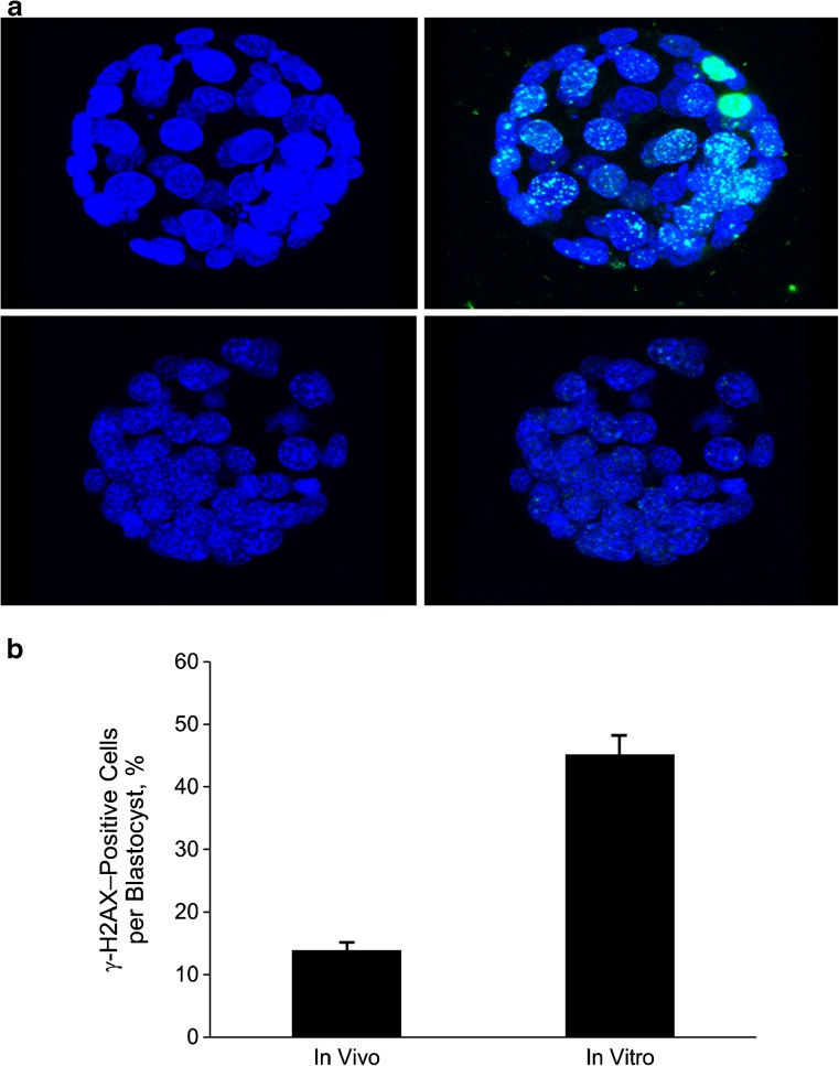
Fig. 2.
A, Upper left: In vitro blastocyst stained with DAPI (blue). Upper right: In vitro blastocyst stained with DAPI and overlaid with γ-H2A.X (green). Lower left; In vivo blastocyst stained with DAPI. Lower right: In vivo blastocyst stained with DAPI and overlaid with γ-H2A.X. B, Percentage of cells in each blastocyst positive for γ-H2A.X (>5 γ-H2A.X foci/nucleus) in the in vivo and in vitro groups (data shown are mean ± standard error; P < .001). DAPI denotes 4’, 6-diamidino-2-phenylindole; γ-H2A.X, phosphorylated histone H2A.X

