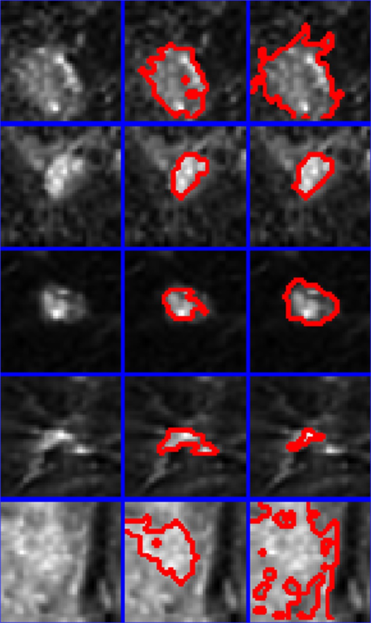
Fig. 3.
Five malignant lesions are presented (left column) along with comparative regions-of-interest produced by the proposed technique (center column) and the 60 % enhancement threshold (right column). Red lines mark the border between a lesion and its surrounding non-suspicious tissue. Blue lines separate pixel patches extracted from examinations included in this study

