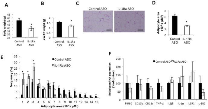
Figure 5. IL-1Ra ASO treatment effect on adipose tissue.
After 6 weeks of treatment with IL-1Ra ASO mice have A) significantly reduced body weight, and B) reduced epididymal fat mass. C) H&E staining of visceral adipose tissue shows reduced adipocyte cell size. Scale bars = 50 µm. Representative figures are presented from the analyses of five different mice per group. D) Quantification of average adipocyte cell area E) Histogram of adipocyte area in control treated mice (black bars), IL-1Ra ASO treated mice (white bars), using Image Gauge software, version 4 (FujiFilm). (n = 5 mice per group), F) QPCR analysis of inflammatory gene expression in adipose tissue. * p<0.05 compared to HFD-fed control treated group, n = 5−8 mice per group.

