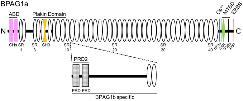
Figure 1. Schematic representation of BPAG1a and b domain organization.
ABD, actin-binding domain; CH, calponin homology domain; SR, spectrin repeat (dotted ovals represent putative SRs, not previously identified as SRs [63], or predicted in mouse or human BPAG1a sequence by SMART [64]; some SRs in the plakin domain were deduced from the alignment with plectin plakin domain [65], SH3, src homology-3 domain (is atypical and embedded in SR5 [66]); PRD, plakin repeat domain; EFhs, EF hands; GAR, GAS2-related domain; GSRs, Gly-Ser-Arg repeats; EBBS, EB1/EB3-binding site containing a Ser-X-Ile-Pro motif (where is X is any residue); MTBD, microtubule-binding domain. BPAG1a (NP_598594) is 5379 res. long. BPAG1b-specific domain (2014 res. long, deduced from NP_604443; 7393 res.) is inserted in between SR10 and SR11 of BPAG1a (after res. 1548).

