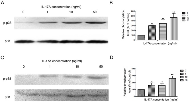
Figure 3. IL-17A activated p38 signaling pathway in NPC cells.
(A) Western blotting analysis was used to detect p38 and p-p38 expression in NPC-039 cells treated with IL-17A (0, 1, 10 and 50 ng/ml) at indicated time points. (B) Quantification of the protein levels of p38 and p-p38 in NPC-039 cells. (C) Western blotting analysis was used to detect p38 and p-p38 expression in NPC-039 cells treated with IL-17A (0, 1, 10 and 50 ng/ml) at indicated time points in CNE-2Z cells. (D) Quantification of the protein levels of p38 and p-p38 in CNE-2Z cells. Values represent the means ± SD of three independent experiments performed in triplicate. *p<0.05 and **p<0.01 compared with the control group.

