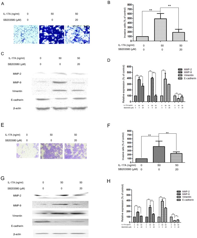
Figure 4. Effects of the p38 inhibitor and IL-17A on cell invasion and MMP-2, MMP-9, Vimentin and E-cadherin expressions in NPC-039 and CNE-2Z cells.
(A) NPC-039 cells were pretreated with SB203580 (20 µM) for 30 min and then incubated in the presence or absence of IL-17A (50 ng/ml) for 24 h. Cellular invasiveness was measured using the transwell invasion assay. (B) The percent invasion rate in NPC-039 cells was expressed as a percentage of control. (C, D) NPC-039 cells were treated and then subjected to western blotting to analyze the protein levels of MMP-2/-9, Vimentin and E-cadherin. (E) CNE-2Z cells were pretreated with SB203580 (20 µM) for 30 min and then incubated in the presence or absence of IL-17A (50 ng/ml) for 24 h. Cellular invasiveness was measured using the transwell invasion assay. (F) The percent invasion rate in CNE-2Z cells was expressed as a percentage of control. (G, H) CNE-2Z cells were treated and then subjected to western blotting to analyze the protein levels of MMP-2/-9, Vimentin and E-cadherin. Values represent the means ± SD of three independent experiments performed in triplicate. *p<0.05 and **p<0.01 compared with the control group.

