
Figure 1. Wound Fluids stimulate growth and self-renewal of tumor initiating cells.
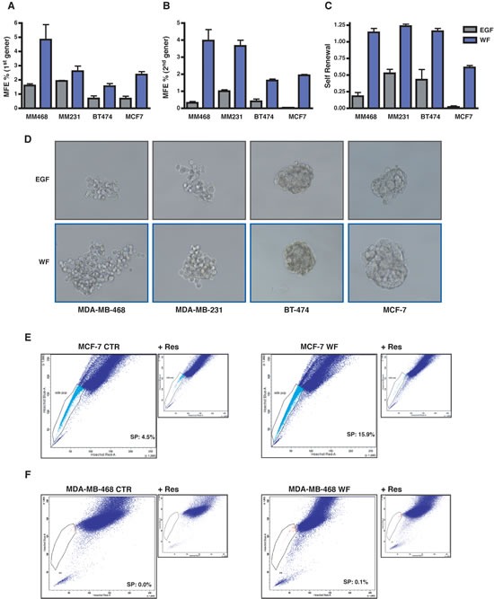
(A) Graph reports primary generation mammosphere forming efficiency (MFE) in MDA-MB-468, MDA-MB-231, BT-474 and MCF-7 cells. Cells were plated as single suspension on poly-HEMA coated dishes in mammosphere standard medium containing EGF or supplemented with 5% wound fluids (WF) and no EGF. MFE was calculated as the ratio between the number of mammospheres and the cells seeded per well. (B) Same as in (A), but on secondary generation mammosphere, i.e. mammospheres formed by cells dissociated and replated as single cells in the indicated medium from primary generation mammospheres. (C) Self-renewal in MDA-MB-468, MDA-MB-231, BT-474 and MCF-7 cells. Self-renewal was calculated as the ratio between numbers of secondary and primary mammospheres. (D) Representative pictures of the mammospheres formed by MDA-MB-468, MDA-MB-231, BT-474 and MCF-7 cells in the primary generation. (E) FACS analysis for evaluation of side population (SP) in MCF-7 cells grown in complete medium (MCF-7 CTR) or in serum free medium supplemented with 5% WF for 48 hours (MCF-7 WF). SP is identified through exclusion of Hoechst dye that is inhibited in the presence of Reserpine. Percentage of SP is reported inside the plot. (F) Same as in (E) but using MDA-MB-468 cells.