
Figure 2. STAT3 is strongly activated in breast cancer cells, following stimulation with Wound Fluids.
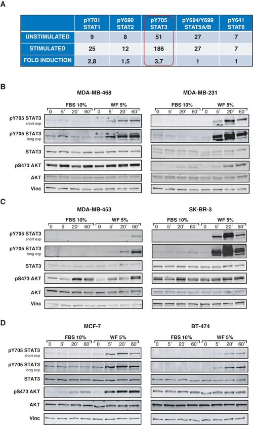
(A) Table reports the activation of five different members of STAT family in MDA-MB-231 cells, following stimulation with 5% wound fluids for 20 minutes (stimulated) or not (unstimulated). Activation was detected using a commercial immunoassay. The value of “fold induction” represents the ratio between the unstimulated and stimulated values. (B) Western blot analysis of MDA-MB-468 and MDA-MB-231 cell lines serum starved and then stimulated for the indicated times with 10% serum (FBS) or 5% wound fluids (WF). (C) Same as in (A), but using MDA-MB-453 and SK-BR-3 cell lines, as indicates. (D) Same as in (A), but using MCF-7 and BT-474 cell lines, as indicated. Vinculin expression was used as loading control.