
Figure 4. Inhibition of STAT3 impacts on growth and self-renewal of tumor-initiating cells.
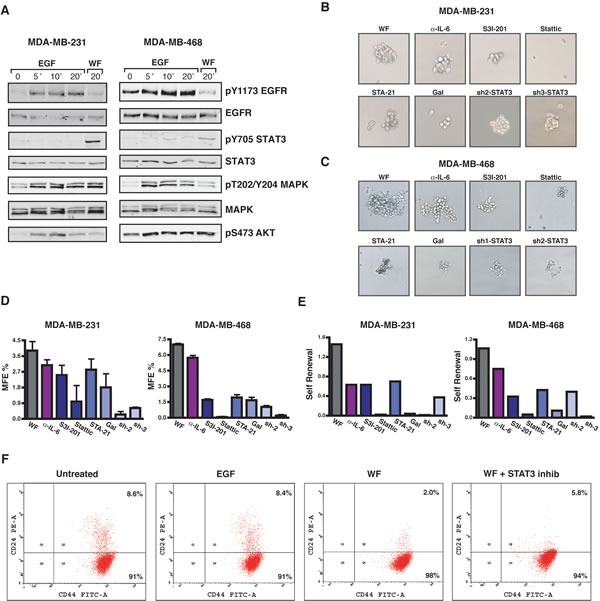
(A) Western blot analysis of MDA-MB-231 and MDA-MB-468 cell lines, serum starved and then stimulated for the indicated times with EGF (20 ng/mL) or 5% wound fluids (WF), as indicated. (B) Images show primary mammospheres formed by MDA-MB-231 cells. Control or STAT3-silenced (sh2 and sh3) cells were plated on poly-HEMA coated dishes in mammosphere growing medium supplemented with 5% wound fluids, in the presence of IL-6 blocking antibody (0.2μg/ml) or STAT3 inhibitors (S3I-201, 50 μM; Stattic, 10 μM; STA-21, 30 μM; and Galiellalactone, 12 μM), as indicated, and grown for ten days. (C) Same as in (B) but using MDA-MB-468 cells. STAT3 inhibitors were used as follows: S3I-201, 100 μM; Stattic, 10 μM; STA-21, 30 μM; Galiellalactone, 25 μM. (D) Graphs report the percent of mammosphere forming efficiency (MFE%) in MDA-MB-231 (left) and MDA-MB-468 (right) cells of the experiment described in (B) and (C). MFE was calculated as the ratio between the numbers of mammospheres counted/number of cells seeded, per well. (E) Graphs report the self-renewal in MDA-MB-231 (left) and MDA-MB-468 (right) cells treated with the inhibitors only during the second generation. Self-renewal was calculated as the ratio between number of secondary mammospheres/number of primary mammospheres. (F) Flow cytometry analysis of CD44highCD24low/neg stem cell-like subpopulation in MDA-MB-231 cells. Percent of CD44highCD24low/neg (Q4) and of CD44highCD24high (Q2) is reported in the plots.