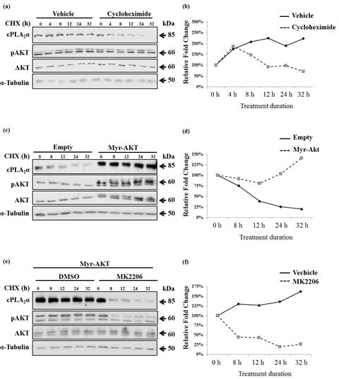
Figure 5. pAKT protects cPLA2α protein from degradation.

(a) PC-3 cells were treated with 50 μM cycloheximide (CHX) for the indicated time. The cells were harvested and subjected to immunoblot analysis. (b) PC-3 cells were transfected with empty vector or Myr-AKT (2 μg, 24 h) followed by incubation with CHX (50 μM) for the indicated time; and (c) PC-3 cells were transfected with Myr-AKT (2 μg, 24 h) followed by incubation with MK2206 (5 μM, 1 h). The cells were then treated with 50 μM CHX for the indicated time. All cPLA2α protein levels were normalized by α-tubulin. The ratio of cPLA2α over α-tubulin at time zero of CHX treatment was set as 100%. AKT antibodies detect AKT1 (62 kDa), AKT2 (56 kDa) and AKT3 (62 kDa, expressed mainly in the brain). When the same concentration of PAGE was used, the separation of AKT 1 from 2 is pending on the running time. The Myr-AKT1 migrates closely with the AKT1 on PAGE.
