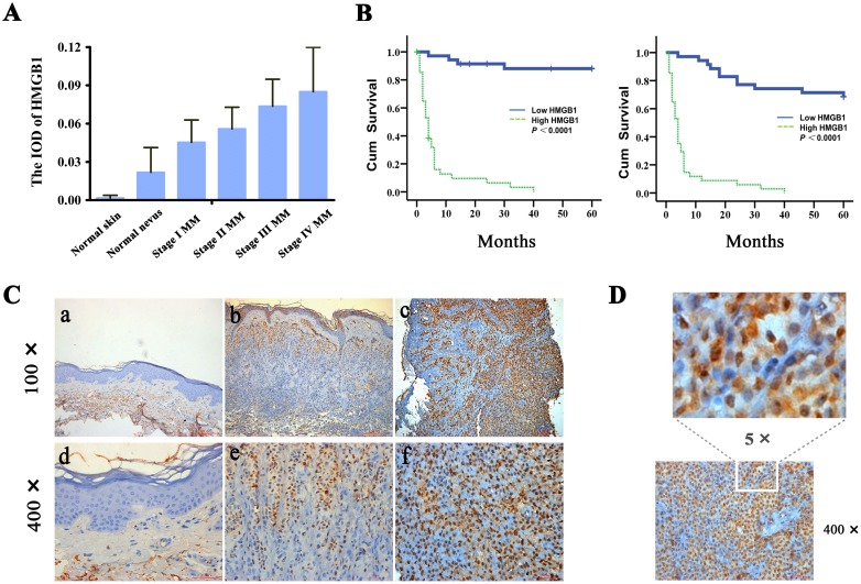
Figure 1. Increased expression of HMGB1 is correlated with the progression of human cutaneous melanoma and poor patient survival.
(A) The expression levels of HMGB1 (IOD) in normal skin, normal nevus, and various melanoma stages (stage I, stage II, stage III and stage IV). TNM classification and clinical stage are according to the melanoma staging system of the American Joint Committee on Cancer (AJCC). The mean IOD of HMGB1 in each group is shown in a bar figure and presented as mean ± SD. As shown, melanomas have increased expression of HMGB1 compared with normal skin and nevi. Low-grade tumors have lower average levels of expression compared with high-grade tumors. Bars: SD; p<0.0001. IOD, integral optical density; MM, malignant melanoma. (B) Kaplan-Meier survival curves for groups based on HMGB1. HMGB1 staining is correlated with overall 5-year survival (left panel), and disease-specific 5-year survival (right panel) (both p<0.0001; log-rank test). Cum,cumulative. (C) Representative immunohistochemical analysis of HMGB1 expression in normal skin (a and d), normal nevi (b and e),and melanoma (c and f). (D) High magnification of HMGB1 detected in melanoma tissue section. As shown, HMGB1 protein was localized mainly inside the melanoma cells.

