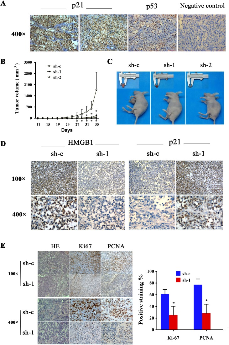
Figure 6. Expression of p21 and p53 in human cutaneous melanoma and effect of HMGB1 inhibition on tumorigenicy of melanoma cell lines.
(A) Representative immunohistochemical staining of p21 and p53 expression in melanoma tissues. MM,malignant melanoma. (B) The A375 cells (5×10⁶/0.2ml) expressing control (sh-c) or HMGB1 shRNA (sh-1 or sh-2) were implanted the flank of nude mouse. Tumor development was monitored 3 times /week. The numbers are means (n=6) of tumor volume ± SD. Bars, SD; *p=0.039. (C) Representative tumor sizes isolated from indicated mice at day 35 post-transplant. (D) Tumors were collected at day 35 post-transplantation. The tumor tissues were processed for immunohistochemical analysis. Representative images of immunohistochemical stain of tumor tissue sections with anti-HMGB1 or p21 antibodies isolated from mice transplanted with melanoma cells expressing sh-c and sh-1. (E) The tumor sections were also stained for H&E, Ki-67 or PCNA. The Ki-67 or PCNA positive counted and the numbers are means from sh-c and sh-1 group. Bars, SD; *p<0.0001.

