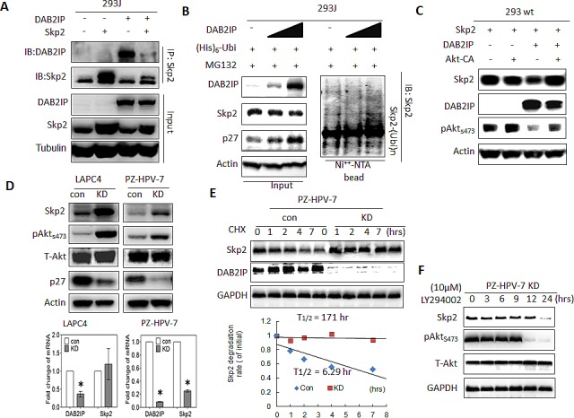
Figure 4. The effect of DAB2IP on Skp2 protein expression mediated through Akt.

(A) 293J cells were transfected with plasmids carrying DAB2IP or Skp2 cDNA for 48 hours. Cell lysates were IP with Skp2 antibody and immunoblotted with DAB2IP or Skp2 antibody (B-C) 293 wt or 293J were transfected with the indicated plasmids. Cell lysates were subjected to western blot, or in vivo ubiquitination assays, respectively. (D) Cell lysates were harvested from control (Con) or DAB2IP knocked-down (KD) cells of LAPC4, PZ-HPV-7 then subjected to western blot and Actin was used as a loading control. Both DAB2IP and Skp2 mRNA expression in LAPC4 KD, PZ-HPV-7 KD and their control cells were determined using qRT-PCR assays. Data are represented as mean +/− SEM. (E) PZ-HPV-7 KD and con cells were treated with cycloheximde (15 μg/ml) at indicated time. Cell lysates were subjected to western blot. The expression of GAPDH was used as a loading control. Skp2 degradation rate was determined based on Skp2/GAPDH ratios at each time point of cycloheximide treatment. (F) PZ-HPV-7 KD cells were treated with 10 M LY294002 at indicated time. Cell lysates were subjected to western blot.
