
Figure 3. sMEK1 and paclitaxel mediate cell migration and cell cycle arrest.
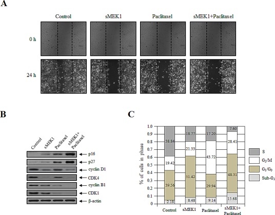
(A) Cell migration potential was monitored using a wound-healing assay. Images of the wound areas were taken at 0 and 24 h using a Leica DFL290 camera. (B) The expression levels of sMEK1, paclitaxel or sMEK1 plus paclitaxel on cell cycle-related proteins, such as p16, p27, cyclin D1, CDK4, cyclin B1 and CDK1 in OVCAR-3 cells. β-actin served as a loading control. (C) Cell cycle progression was evaluated using FACScalibur; data are presented as characteristic DNA histograms. Cells were treated with FITC-labeled Annexin V, PI (Boehringer Mannheim, Mannheim), and RNase A (1 mg/ml) in PBS, and then incubated for 1 h at 37°C in the dark.