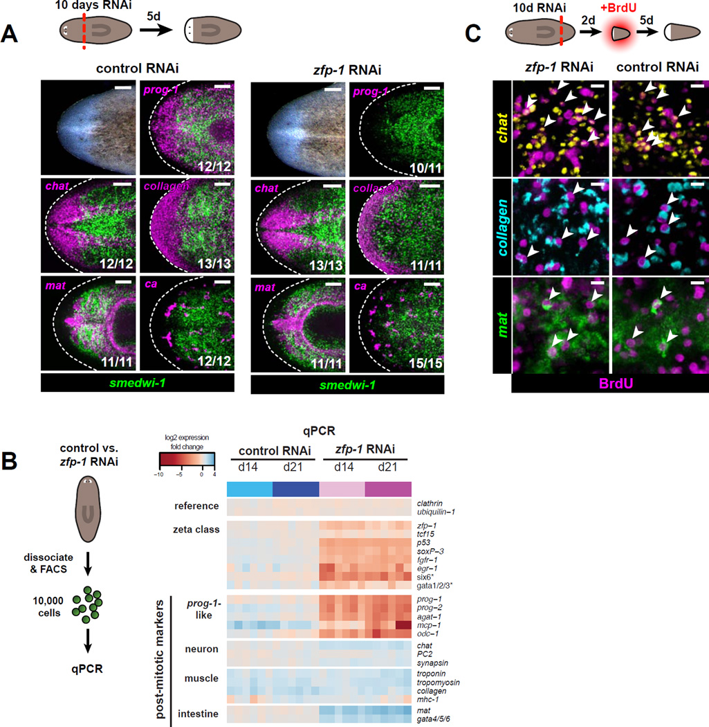
Figure 5. Neoblasts maintain the ability to form blastemas and regenerate multiple tissue types in the absence of the ζ-class.
A. Regenerated head blastemas from 10-day RNAi animals were analyzed 5 days post-amputation. Tissues are labeled by RNA probes for ca (nephridia), chat (CNS), collagen (muscle), mat (intestine), prog-1, and smedwi-1 (neoblasts). Numbers of animals with displayed tissue patterns are indicated. Dotted lines indicate animal boundary. Anterior, left. Scale bars, 100 µm.
B. Transcriptional changes in ζ-class transcripts and post-mitotic tissue markers following zfp-1 RNAi, analyzed by qPCR. Biological replicates (10,000 live cells each) were isolated by FACS after control RNAi (blue) or zfp-1 RNAi (magenta). Heatmap shows log2 fold changes in expression levels (relative to day 0) after global median normalization.
C. Tail fragments cut from 10-day RNAi animals were soaked in BrdU starting 2 days post-amputation and fixed 5 days later. Specimens were assessed by triple FISH for chat (CNS), collagen (muscle), and mat (intestine), together with BrdU IF. Representative confocal projections through head blastemas regenerated following zfp-1 or control RNAi are shown. BrdU-positive cells expressing each tissue marker are indicated by arrowheads. Scale bars, 10 µm.

