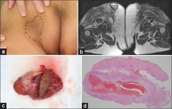
Figure 1.

(a) A well-circumscribed subcutaneous mass, measuring 6 cm, on patient's left buttock; (b) T2-weighted magnetic resonance imaging. A fluid–fluid level was clearly observed; (c) the resected mass. A cystic mass with pseudocapsule; (d) histopathological findings (hematoxylin and eosin, low magnification). A pseudocapsule containing red blood cells with a fibrous wall
