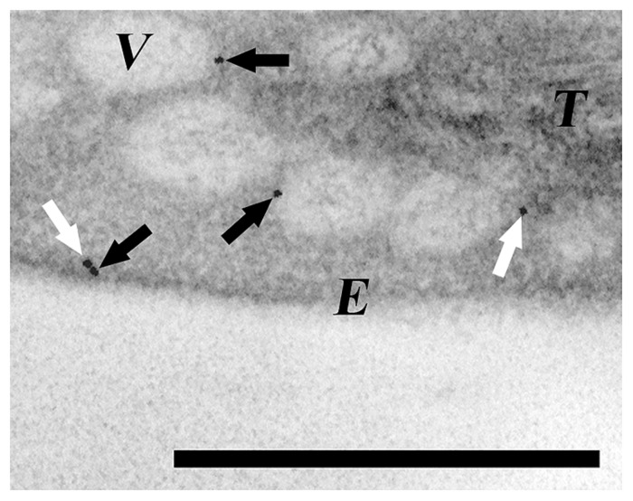
FIGURE 1.
Detection of CPSAR1 in Arabidopsis chloroplasts. Transmission electron micrograph of immunogold-labeled sections of leaves after low temperature incubation (Garcia et al., 2010) showing the presence of CPSAR1 in the stroma (white arrow) and at both the envelope and vesicle membranes (black arrows). E, envelope; V, vesicle; T, thylakoids. Bar 0.5 μm.

