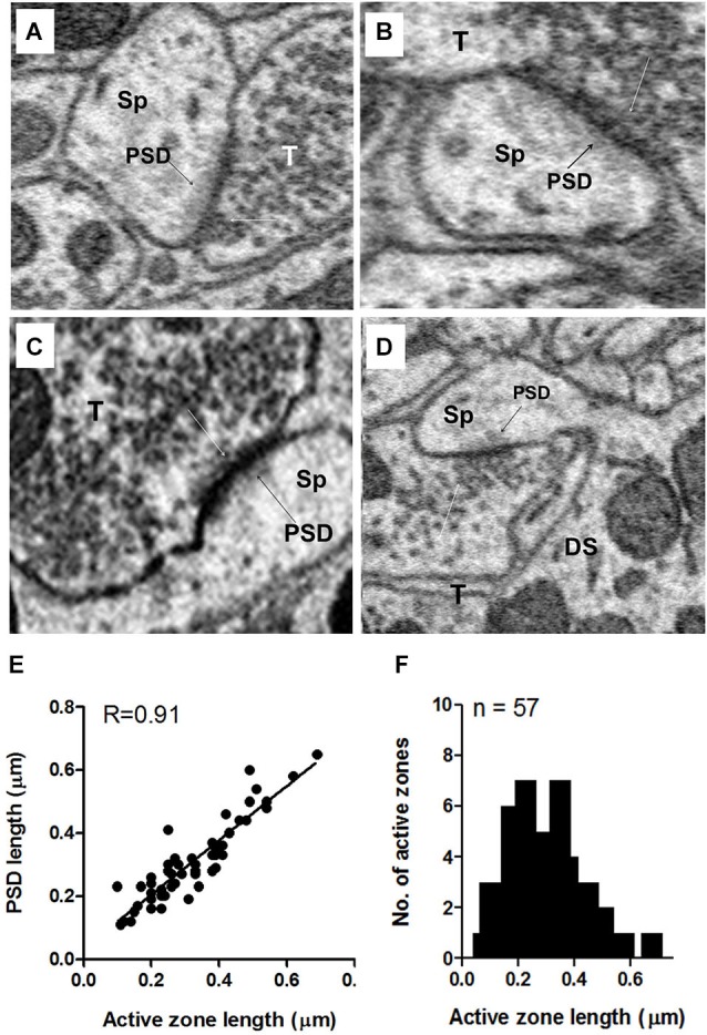
Figure 10.

Examples of axo-spinous synapses on apical dendrites showing active zones and postsynaptic densities (PSDs). (A–D) The axon terminal (T) abutting the spine (Sp) contained a single active zone with an associated cluster of docked vesicles (white arrows). The PSD appears as a dark shadow beneath the contact with the presynaptic terminal (T) of the spine. As in the terminals on the soma, the vesicle cluster associated with the active zone was an extension of a larger pool of vesicles that spread throughout and often filled much of the terminal. (E) Relationship between the active zone and PSD lengths of axo-spinous synapses. (F) Maximum length distribution of active zones of axo-spinous synapses. Data for (E) and (F) were obtained from a random sample of 57 synapses from the apical dendrites of three fusiform cells.
