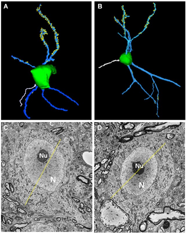
Figure 2.

Morphological features of fusiform cells. (A,B) 3D-reconstructions of the cell somata (green) and selected apical dendrites (blue) containing spines (yellow). An axon (white) can be seen extending from the soma of each neuron. (C,D) Transverse sections through each fusiform cell shown in (A,B) at levels where they reached their largest diameters (yellow lines). The diameters were 26.38 μm for the fusiform cell shown on the left and 24.4 μm for the other cell. N: Nucleus, nu: nucleolus.
