
Figure 2.
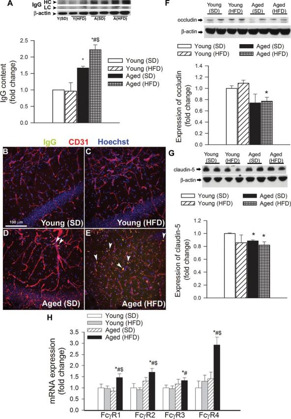
Obesity in aging exacerbates disruption of the blood–brain barrier. (A) Obesity and aging-induced changes in immunoglobulin G (IgG) content in the hippocampus of mice. (Upper panel) Original Western blot showing IgG heavy chain (HC) and light chain (LC) expression. β-Actin was used as a loading control. Bar graphs are summary densitometric values. Data are mean ± SEM. *p < .05 vs Young (standard diet [SD]); # p < .05 vs Young (high-fat diet [HFD]); $ p < .05 vs Aged (SD). (B–E) Confocal microscopy analysis of plasma-derived IgG (green) and CD31-positive microvessels (red) in the hippocampus of young and aged mice with or without HFD-induced obesity. Note the increased presence of extravascular IgG deposits in the hippocampus of aged obese mice (arrowheads). (F and G) Expression of the tight junction proteins occludin (F) and claudin-5 (G) in the hippocampi of young and aged SD-fed lean and HFD-fed obese mice. (Upper panels) Original Western blots. β-Actin was used as a loading control. Bar graphs are summary densitometric values. Data are mean ± SEM. *p < .05 vs Young (SD). n = 4–6 animals per group. (H) Quantitative reverse transcription–PCR data showing mRNA expression of the Fc-gamma receptor (FcγR) isoforms in the hippocampi of young and aged SD-fed lean and HFD-fed obese mice. Data are mean ± SEM (n = 6 in each group), *p < .05 vs Young (SD); # p < .05 vs Young (HFD); $ p < .05 vs Aged (SD).