Figure 3.
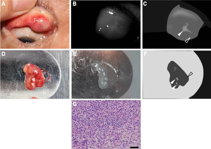
Case 5: a 61-year-old man with chalazion.
Notes: (A) Photograph of the tarsal conjunctiva of the right upper eyelid. A subconjunctival mass was observed at the nasal side. (B) Noninvasive meibographic image of the tarsal conjunctiva of the right upper eyelid and (C) its schematic representation. The mass showed overall low reflectivity (open arrowhead) but contained a central region of high reflectivity (closed arrowhead). (D) Photograph of the curettage specimen including fatty granules. (E) Noninvasive meibographic image of the curettage specimen and (F) its schematic representation. Noninvasive meibography detected overall low reflectivity of the granuloma lesion (open arrowhead) but high reflectivity of fatty granules (closed arrowheads). (G) Histopathologic analysis of the granuloma lesion. Inflammatory cells and dense fatty vacuoles were observed (hematoxylin–eosin; bar =100 μm).

