Figure 6.
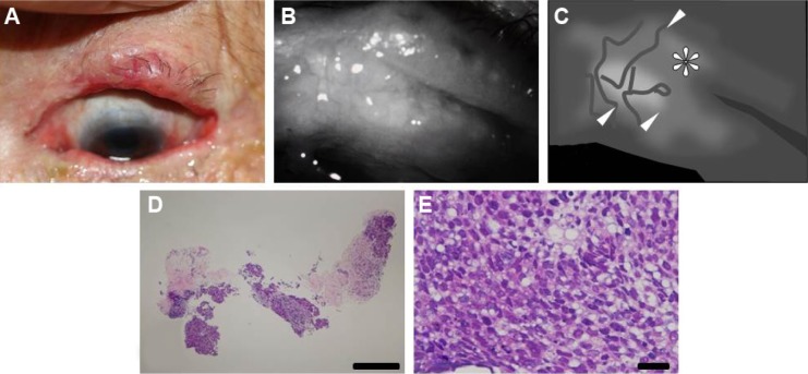
Case 8: an 82-year-old woman with recurrent sebaceous carcinoma.
Notes: (A) Photograph of the left upper eyelid. A nodular lesion with an irregular edge was detected. (B) Noninvasive meibographic image of the tarsal conjunctiva of the left upper eyelid and (C) its schematic representation. Noninvasive meibography revealed a poorly bordered lesion with relatively high intensity in both the nodular region and tarsal conjunctiva (arrowheads). The nodular region of the incisional biopsy is indicated (asterisk). Histopathologic analysis of the incisional specimen from the nodular lesion shown at (D) lower and (E) higher magnification. The nodular region was invaded by tumor (hematoxylin–eosin; bar =1 mm). (E) Neoplastic cells with fatty vacuoles were sparsely distributed in the specimen (hematoxylin–eosin; bar =50 μm).

