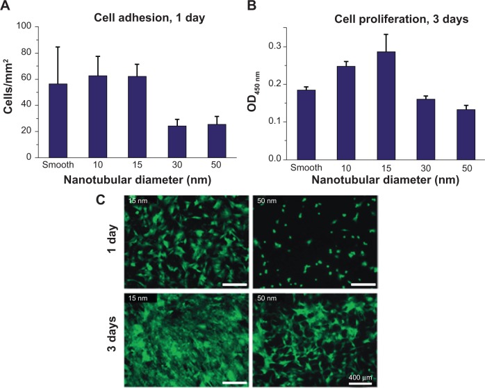
Figure 6.
Cell densities of adherent cells on ZrO2 nanotubes with different diameter count under fluorescence microscope after 24 hours’ adhesion (A) and measured using colorimetric WST-assay after 3 days’ proliferation (B). Fluorescence images of GFP-labeled mesenchymal stem cells after 24 hours’ adhesion and 3 days’ proliferation (C).
Note: Copyright © 2009. Reproduced by permission of the Royal Society of Chemistry, from Bauer S, Park J, Faltenbacher J, Berger S, von der Mark K, Schmuki P. Size selective behavior of mesenchymal stem cells on ZrO2 and TiO2 nanotube arrays. Integr Biol (Camb). 2009;1(8–9):525–532.81
Abbreviations: GFP, green fluorescence protein; OD, optical density; WST, water soluble tetrazolium salts.

