Figure 1.
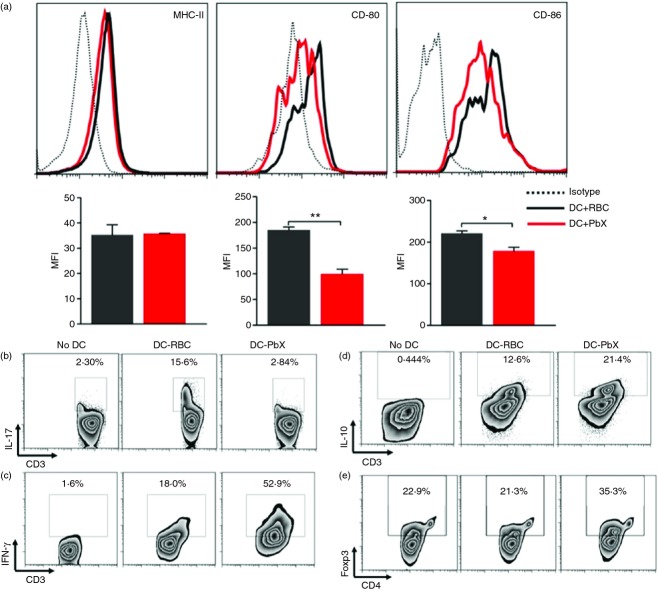
Dendritic cells (DCs) treated with Plasmodium berghei extracts (PbX) acquire an immature phenotype and stimulate the generation of regulatory T cells. Bone marrow-derived DCs were stimulated with lipopolysaccharide (LPS; 1 µg/ml) for 18 hr in the presence of PbX (100 μg/ml). As control, cells were stimulated in the presence of non-infected red blood cell extracts (RBC, 100 μg/ml). (a) The cells were collected and stained with fluorochrome-conjugated antibodies for MHC-II, CD80 and CD86. The mean fluorescence intensity (MFI) was calculated from 500 000 acquired events. Dendritic cells were treated with PbX (100 μg/ml) or non-infected RBC extracts (100 μg/ml), stimulated with LPS (1 µg/ml) and pulsed with myelin oligodendrocyte glycoprotein peptide (MOG35–55) peptide (10 μg/ml) for 18 hr. Then cells were seeded in 96-well U-bottom plates (5 × 10⁵ cells/well). T lymphocytes were isolated from spleens of naive mice using Dynabeads and seeded with DCs (1 DC : 1 T). The cells were allowed to grow for 96 hr in the presence or absence of MOG peptide (10 μg/ml). As controls, T cells were cultivated without DCs. At the end of the culture period, the supernatant was collected and the cells were treated with PMA and Ionomycin together with Brefeldin-A for 4 hr. (b–d) Intracellular interleukin-17 (IL-17), interferon-γ (IFN-γ) and IL-10 in CD3+ cells was determined, respectively. (e) The generation of Foxp3+ regulatory T cells was also determined. Data from three separate experiments with similar results. *P < 0·05 and **P < 0·01.

