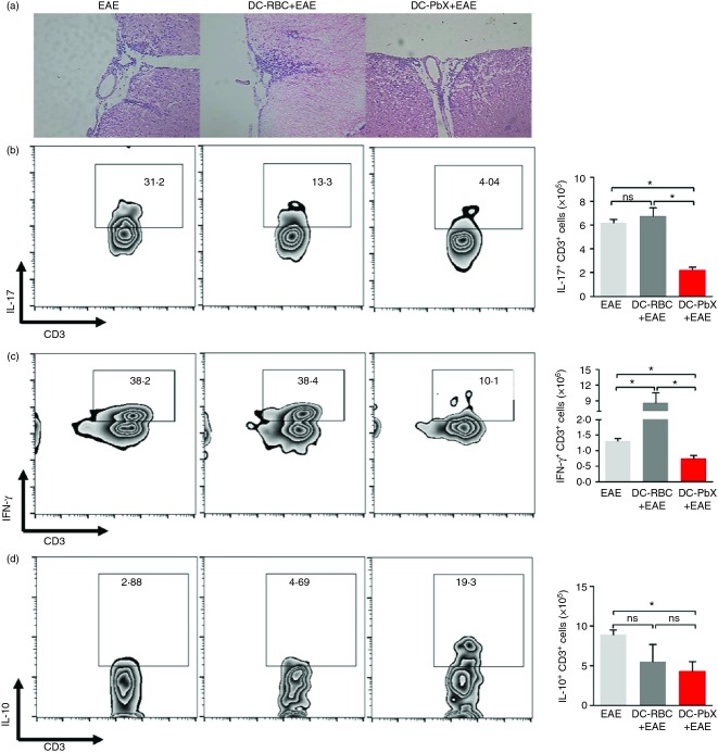
Figure 3.
Changes of infiltrating cells in the central nervous system (CNS) from dendritic cell (DC) -transferred mice. DCs were stimulated with lipopolysaccharide (LPS; 1 µg/ml) then treated with Plasmodium berghei extracts (PbX; 100 μg/ml) and pulsed with myelin oligodendrocyte glycoprotein peptide (MOG35–55) peptide (10 μg/ml) for 18 hr and adoptively transferred (1·5 × 106 cells/mouse) into C57BL/6 mice (n = 6/group) before the induction of experimental autoimmune encephalomyelitis (EAE). (a) At day 14 after immunization, the CNS tissue of EAE-effected mice was removed and processed for histopathology analysis by haematoxylin & eosin staining. Magnification: 200×. In addition, the CNS tissue of EAE mice (both DC-transferred and untreated) was removed at day 14 post-immunization and the infiltrating leucocytes were enriched through gradient centrifugation using Percol reagent. Cells were stimulated with PMA + ionomycin in the presence of Brefeldin-A for 4 hr before staining with fluorochrome-conjugated antibodies. The frequencies of T cells producing interleukin-17 (IL-17) (b), interferon-γ (IFN-γ) (c) and IL-10 (d) were evaluated by flow cytometry. Data from three independent experiments. *P < 0·05.

