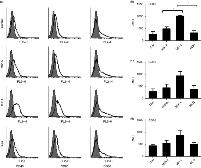
Figure 2.
Expression of activation markers by macrophages stimulated with heat-killed Mycobacterium indicus pranii (MIP-K), live Mycobacterium indicus pranii (MIP-L) and bacillus Calmette–Guérin (BCG). RAW 264.7 macrophages were stimulated with MIP-K, MIP-L and BCG at a multiplicity of stimulation of 10. Cells were stained with phycoerythrin-conjugated anti-mouse CD40, CD80 and CD86 monoclonal antibodies and analysed by flow cytometry. MIP-L was leading to higher up-regulation of co-stimulatory molecules. Moderate up-regulation was observed in response to MIP-K. Representative histograms are shown. Composite Mean Fluorescence Intensity (= % positive cells × mean fluorescence intensity of positive cells) is also shown as a bar graph. Data shown are mean ± SEM of two independent experiments. *P < 0·05.

