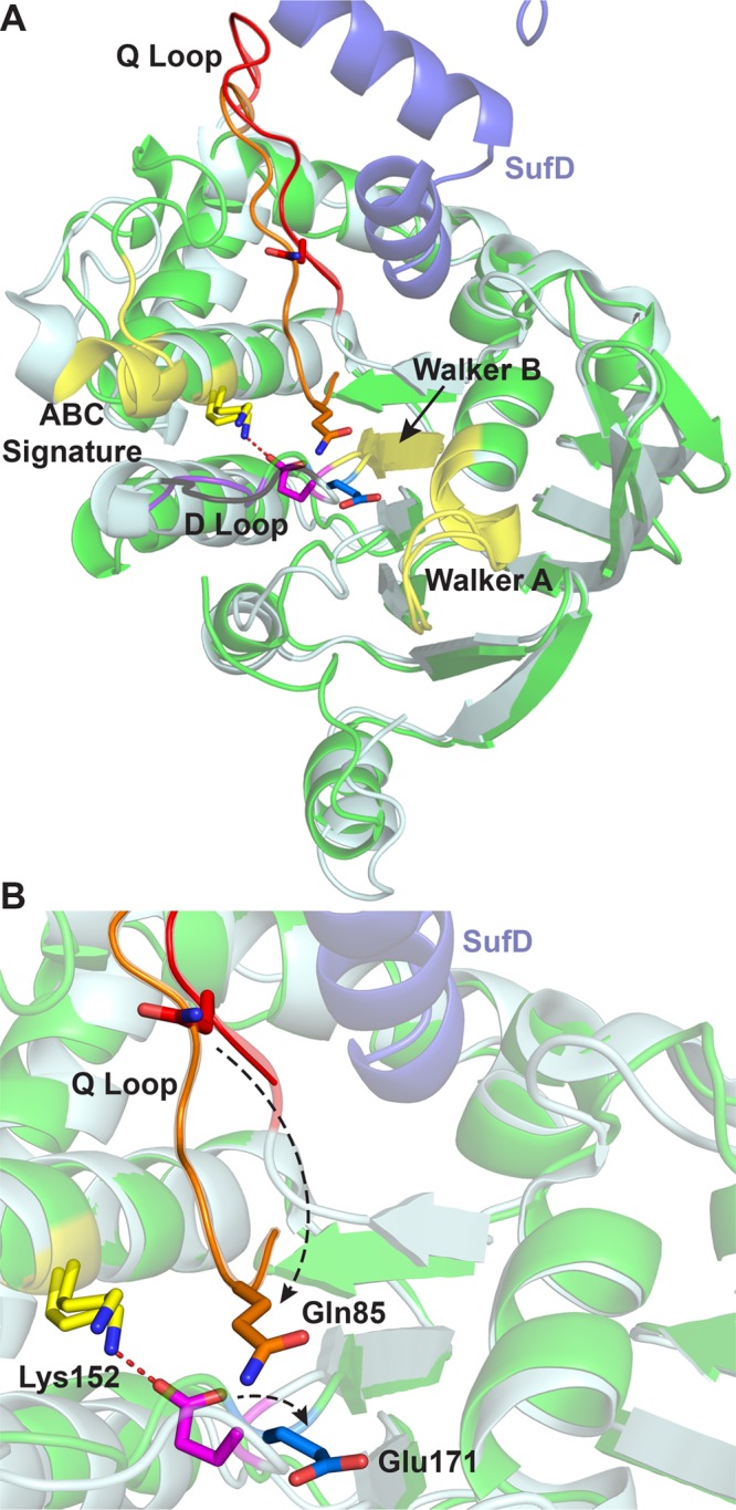
Figure 3.

(A) Structural alignment of the nucleotide-free SufC monomer (light blue, PDB entry 2D3W) and one nucleotide-free SufC monomer from the SufC2D2 complex (green, chain C of PDB entry 2ZU0). C-Terminal helices 6 and 7 of SufD from SufC2D2 are shown at the top (blue). The Walker A, Walker B, and ABC signature motifs are colored yellow. The Q-loop is colored red (SufC monomer) or orange (SufC2D2). The D-loop is colored black (SufC monomer) or purple (SufC2D2). (B) Close-up view of panel A. Black dashed arrows indicate positional changes of those residues going from the structure of SufC alone to the SufC from SufC2D2. Gln85 (red to orange), Lys152 (yellow), and Glu171 (pink to cyan) are shown as sticks, and the Lys152–Glu171 salt bridge is shown as a red dashed line.
