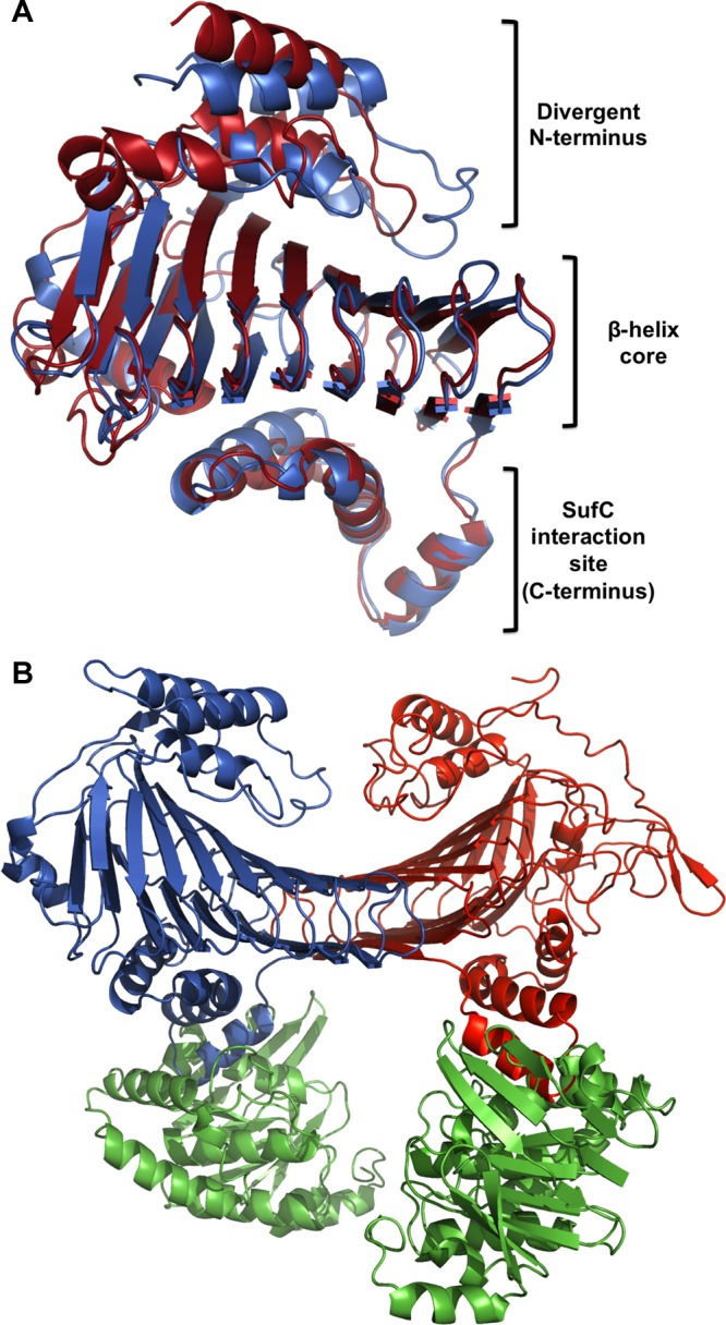
Figure 4.

(A) Structural alignment of E. coli SufD (PDB entry 1VH4) and Methanosarcina mazei Go1 SufB (PDB entry 4DN7). The alignment was generated using the FATCAT Pairwise Alignment tool. (B) Model structure of the E. coli SufBC2D complex generated by modeling E. coli SufB on one chain of the SufC2D2 structure (PDB entry 2ZU0). SufB is colored red; SufC monomers are colored green, and SufD is colored blue. The alignment was generated using the FATCAT Pairwise Alignment tool.
