Figure 6.

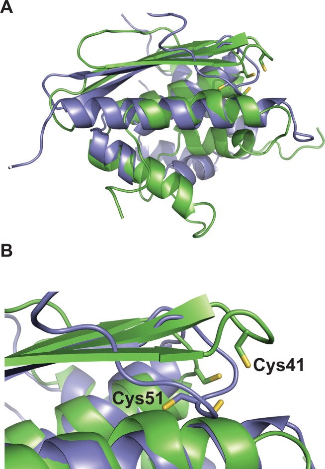
(A) Structural alignment of E. coli SufE (purple, PDB entry 1MZG) and B. subtilis SufU (green, PDB entry 2AZH). Active site Cys residues are show as sticks. (B) Enlarged view of the alignment showing the relative orientation of SufE Cys51 and SufU Cys41. The alignment was generated using the FATCAT Pairwise Alignment tool.
