Figure 3.
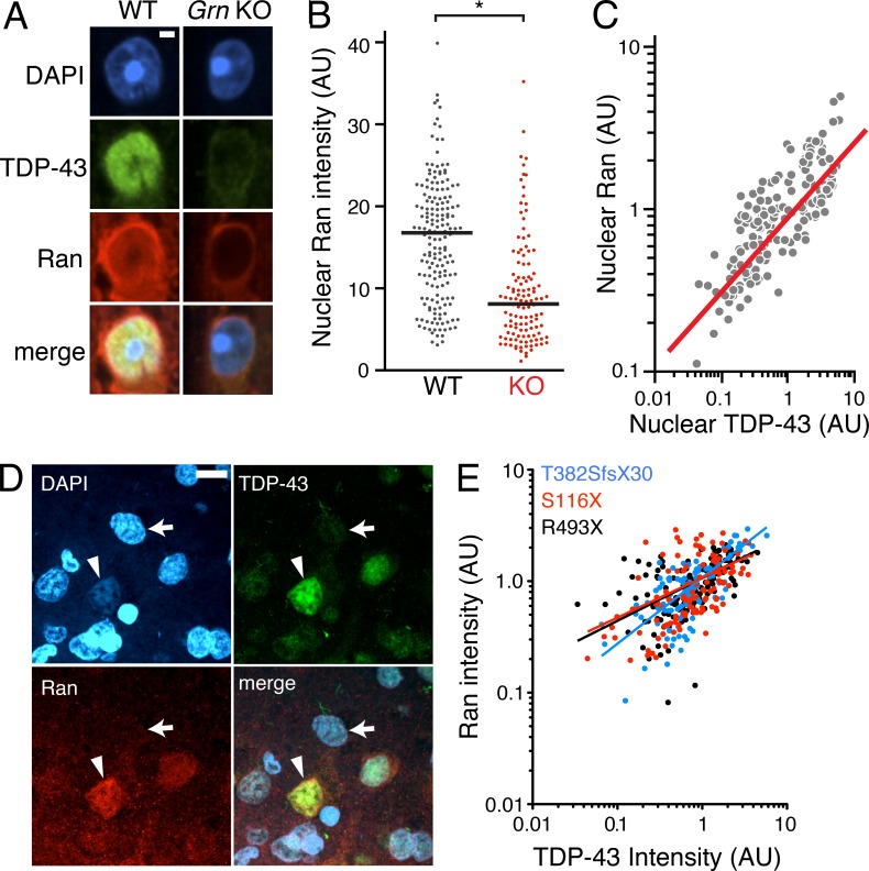
Nuclear clearing of TDP-43 and Ran are pathologically associated in FTLD-TDP. (A) 18-mo-old GCL neurons from WT and Grn-KO retinas were co-stained for TDP-43 and Ran. Nuclei were labeled with DAPI. (B) Nuclear Ran levels in 18-mo-old GCL neurons. n = 165–278 cells from 6 mice/genotype; *, P = 0.019, linear regression model; 2 independent experiments. Scatter plot of individual cell intensities with medians shown. (C) Nuclear Ran and TDP-43 intensities are correlated in Grn-KO GCL neurons. Each dot represents a single cell. n = 165 cells from 6 Grn-KO mice; r = 0.8963; P < 0.001, Spearman’s rho; 2 independent experiments. (D) Immunofluorescence co-staining of GRN mutant human cortex shows depletion of Ran and TDP-43 in the same neuron (noted with an arrow; compare to neurons with high levels of TDP-43 and Ran [arrowhead]). (E) TDP-43 and Ran levels correlate in cortical neurons from human GRN-mutation carriers. Shown are the correlation analyses of nuclear Ran and TDP-43 intensities of individual neurons from post-mortem brain. n = 111–141 cells from each of 3 subjects;, r = 0.56; P < 0.001. The serum progranulin levels were 19.3–21.2 ng/ml for R493X carrier (control patients: 41.3 ±15.5 ng/ml). Spearman’s rho. Bars: 2 µm (A), 10 µm (D).

