


FIG 2.
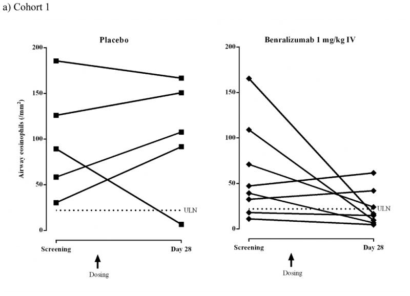
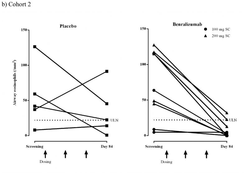

Airway eosinophils (/mm2) for individual subjects (ATP population).
a) Cohort 1 IV: placebo, n = 5; benralizumab 1 mg/kg, n = 8.
b) Cohort 2 SC: placebo, n = 5; benralizumab 100 mg, n = 4; benralizumab 200 mg, n = 5. Dashed lines represent upper limit of normal (ULN) for airway eosinophil counts (22/mm2).4,15
c) Cohort 2 SC. Representative immunofluorescence photomicrographs showing mucosal and submucosal tissue sections obtained from one placebo subject and one benralizumab subject at screening and day 84. Green indicates anti-major basic protein-1-stained eosinophils and blue indicates 4′,6-diamidino-2-phenylindole (DAPI)-stained nuclei. The inserts show sections at x400 magnification.