
Figure 2. Optic glioma-associated vision disturbances are greater in girls with NF1 and in female Nf1-CKO mice.
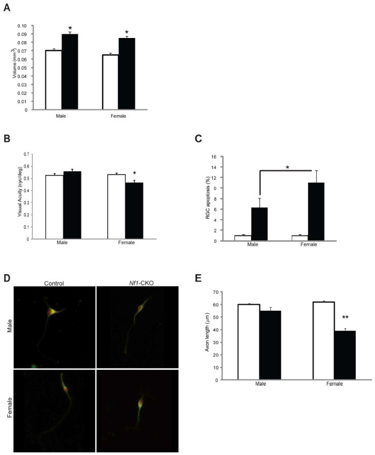
(A) Optic nerve volumes in Nf1-CKO mice (black bars) are larger than those observed in control littermate mice (white bars), regardless of sex. Asterisks denotes p<0.05. (B) Only female Nf1-CKO mice have impaired visual acuity (VOS; cycles/degree) relative to controls. (C) Greater retinal ganglion cell apoptosis (%TUNEL+ cells) was observed in female Nf1-CKO mice (11.5-fold over female controls) relative to Nf1-CKO males (6.2-fold over male controls). (D) Representative images of retinal ganglion cells in culture demonstrate that female, but not male, Nf1-CKO neurons have reduced axon lengths relative to controls. (E) Female, but not male, Nf1-CKO neurons exhibit shorter axon lengths (~50% reduction) relative to controls. Open (white) bars denote control (CTL) mice; closed (black) bars denote Nf1-CKO mice. *p<0.05, **p<0.01.