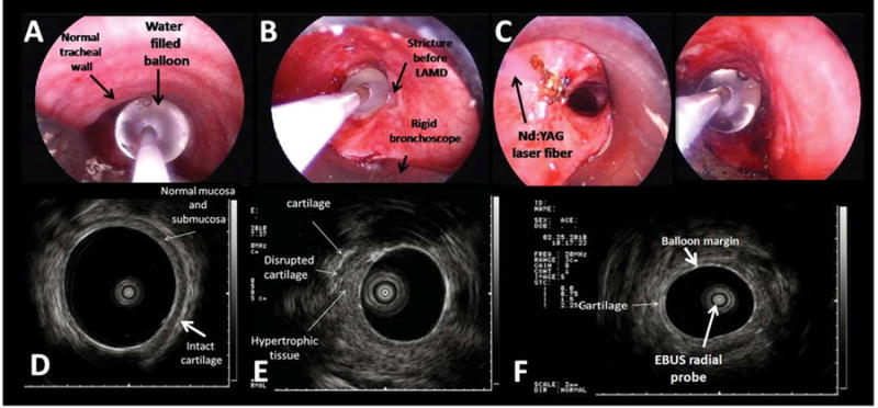
Fig. 5.

(A) WLB image shows the radial EBUS probe with the water-filled balloon overlying the normal tracheal wall. (B) The water-filled balloon completely occludes the airway lumen at the level of the stricture before laser and dilation. (C) WLB view of Nd:YAG laser fiber and water-filled balloon of the radial EBUS probe at the level of the left hypertrophic tissue. (D) EBUS image of the normal tracheal wall. The submucosa and the cartilaginous layers are clearly identified overlying the water-filled balloon. (E) EBUS image of the stricture before laser and dilation at the level of the left hypertrophic tissue. The thick layer of the hypertrophic tissue is visualized overlying a portion of disrupted cartilage. (F) EBUS image of the stricture after laser and dilation shows the water filled balloon overlying the cartilage.
