
Figure 4. Bax is an essential for SMBA induction of apoptosis and Cyt c release.
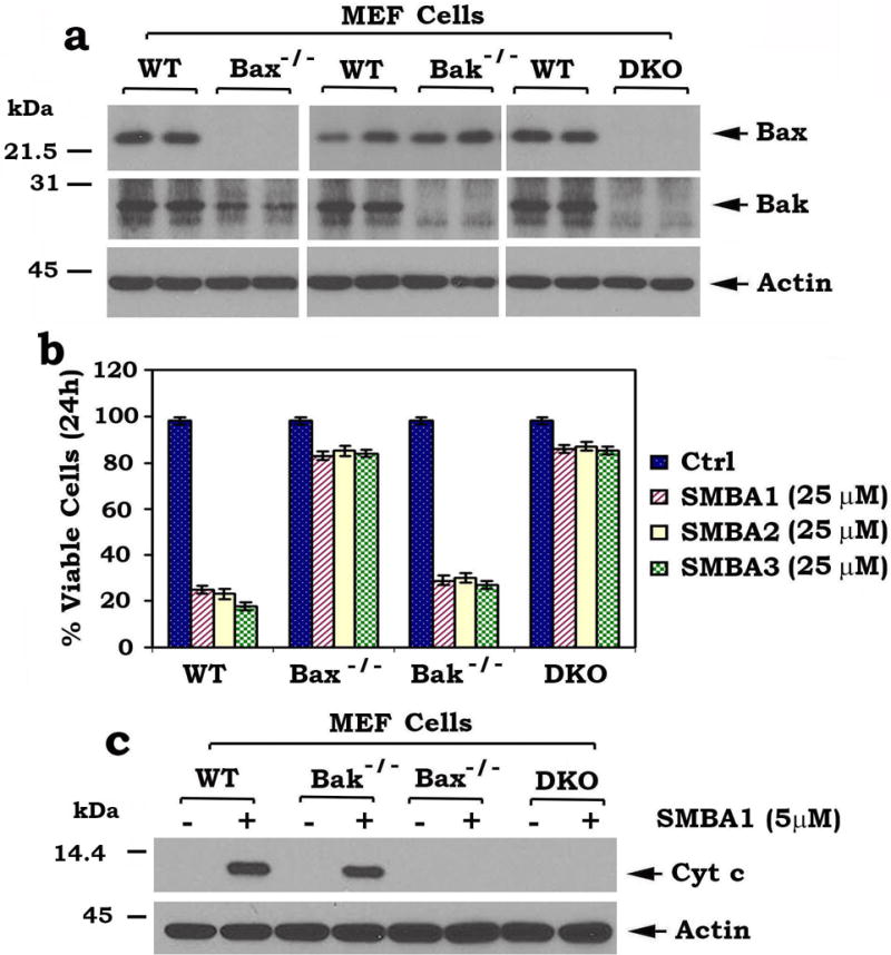
(a) Levels of Bax were analyzed by Western blot in wild type (WT), Bax−/−, Bak−/− or Bax/Bak double knockout (DKO) MEF cells. (b) WT, Bax−/−, Bak−/− or DKO MEF cells were treated with high concentration (25 μM) of SMBA 1, 2 or 3 for 24h. Cell viability was analyzed by PI- Annexin-V staining with FACS analysis. The error bars indicate ± SD of three separate experiments. (c) Mitochondria were isolated from wild type (WT), Bax−/−, Bak−/− or DKO MEF cells. The isolated mitochondria were then treated with lead compound SMBA1 (5μM) in mitochondrial bufferfor 30 min at 30°C. After centrifugation, Cyt c in supernatant fraction (i.e. Cyt c release) and actin in mitochondrial fraction were analyzed by Western blot (full blots can be found in Supplementary Fig. 8).