
Figure 1. MTS assay to determine functional dose range of EISO in HaCaT cells.
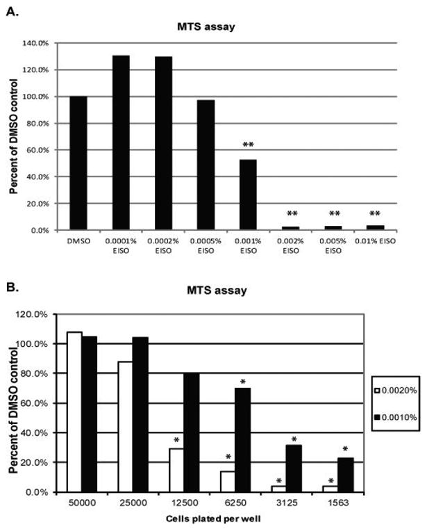
(A) HaCaT keratinocytes grown in a 96-well plate were serum-starved for 24 hr and then treated with a range of EISO from 0.0001-0.01% diluted in DMSO in triplicate for a period of 24 hr. (B) HaCaT cells were plated at varying cell densities so that the initial number of cells per well ranged from 1,563 to 50,000 cells using serial 1:1 dilutions of cells into DMEM. Again, cells were serum starved for 24 hr before treatment with 0.001% or 0.002% EISO in triplicate for an additional 24 hr. In all experiments, cell growth medium was spiked with MTS substrate for a period of 15 minutes and then an OD was obtained using a wavelength of 490 nm. Triplicate values were averaged and means expressed as a percentage of the reading for DMSO-treated control cells. In (A), triplicate wells were treated with DMSO, and in (B) a triplicate cell control was run for each density plated.