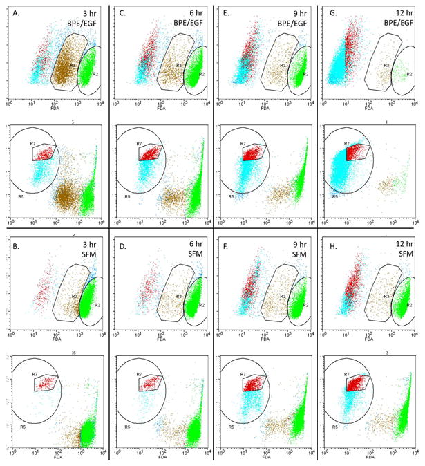
Figure 4. Proliferating HaCaT cells were more sensitive to EISO-induced plasma membrane damage than quiescent cells.

HaCaT cells were starved in serum-free DMEM for 24 hr. Fresh media supplemented with bovine pituitary extract and epidermal growth factor (A, C, E, G) or not supplemented (B, D, F, H) was added to the cells for 3 hr prior to treatment with EISO. At the indicated times, cells were detached by trypsinization and pooled with floating cells. Cells were pelleted, resuspended in PBS and incubated with fluorescein diacetate and propidium iodide for 15 minutes before analysis by flow cytometry. Using control cells loaded with individual dyes, regions of cells were identified as live cells with uncompromised membranes (R2), live cells with compromised membranes (R3), or severely compromised cells. Serum-starved EISO-treated cells (6 hr; panel D) displayed 2 distinct PI-stained populations of actively cycling cells in G1 and G2 phases (R7) that was used to confirm the population of cells with compromised membranes (evidenced by reduced fluorescein retention) but that were still alive.
