Figure 3.
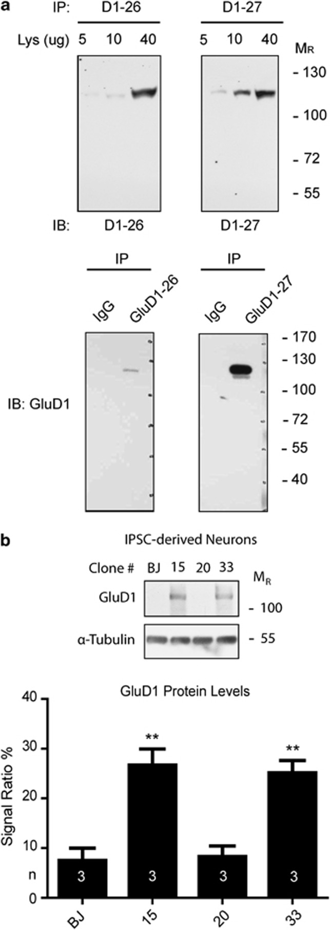
(a) Anti-GluD1 antibody characterization. Different amounts of mouse brain lysate as well as immunoprecipitates (with mouse nonspecific IgG as control) were loaded and probed for two different antibody batches (26 and 27) to double-proof the identity of the recognized epitope. (b) GluD1 expression in iPSC-derived neurons. Each lane was loaded with 30 μg of total protein. Experiments were performed in triplicate (n=3). Immunosignals were detected using autoradiographic films, and glutamate-δ-1 receptor levels were quantified using Photoshop CS3 software (Adobe Systems, San Jose, CA, USA) and normalized against α-tubulin levels (**P<0.01, Student's t-test).

