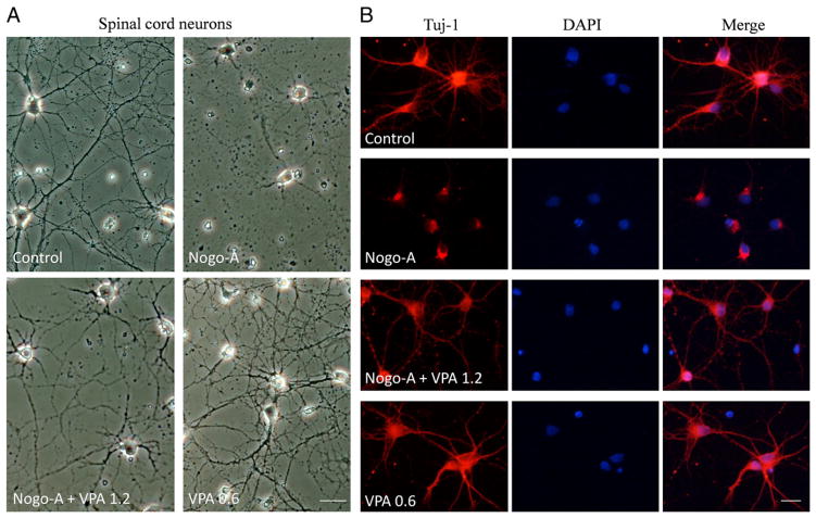
Fig. 4.
VPA attenuated Nogo-A inhibition on axonal growth of spinal neurons. (A) Spinal neurons were cultured on 24-well coated with or without Nogo-A peptide. Compared to control, Nogo-A inhibited axonal growth. However, VPA exposure attenuated Nogo-A inhibition and promoted neurite outgrowth. (B) Neurons were immunostained with antibodies to β-III tubulin to trace neurite (red), and nuclei were stained by DAPI (blue). VPA obviously enhanced neurite generation. Bar A=40 μm, Bar B=100 μm.

