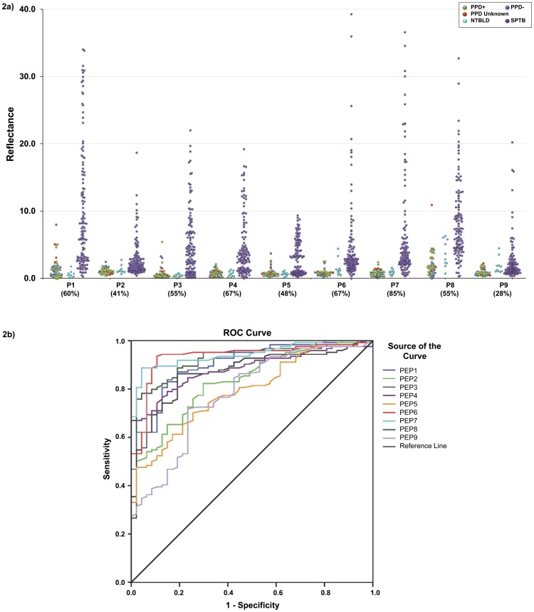
Figure 2. Reactivity of sera with individual BSA-peptide conjugates in lateral flow format.
(A) Dot plot of reflectance values obtained with sera from healthy subjects [PPD- (blue), PPD+ (light green), PPD unknown (red); n = 47], non-TB lung disease patients [NTBLD (light blue); n = 10], and sputum smear-positive TB patients [SPTB (purple); n = 124, including 14 patients co-infected with HIV] tested for reactivity to BSA-peptide conjugates on lateral flow are plotted. Dashed lines indicate mean reflectance of the 47 healthy subjects plus 2.5 times the standard deviation which was used as cut-off to determine sensitivity in TB patients. Values in parenthesis under dot-plot for each BSA-peptide represent percent sensitivity of anti-peptide antibody detection obtained in TB patients with that peptide. The difference between healthy controls and TB patients was highly statistically significant (P<0.0001) for all 9 peptides. The P values for difference between the NTBLD and TB patients ranged from 0.0073−<0.0001 for P1-P8; for P9 the P value was 0.7221. (B) ROC curves representing the performance of the 9 BSA-peptide conjugates (P1–P9) on lateral flow format for reactivity with sera from healthy subjects and smear-positive TB patients.

