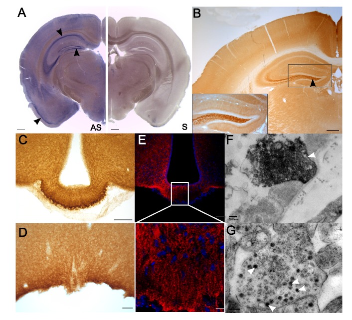
Figure 2. Rbcn-3α is expressed in exocytosis vesicles in the external layer of the median eminence.
(A) ISH with a mouse Dmxl2 antisense probe (AS) and a sense probe (S). (B) Immunolabeling with an antibody against Rbcn-3α revealed high levels of Dmxl2/Rbcn-3α expression in the dentate gyrus, the CA1 and CA3 regions of the hippocampus, and the cortex (black arrowheads). Scale bars, 200 µm. (C and D) Rbcn-3α was found to be strongly expressed in the external layer of the ME (C) and the OVLT (D). Scale bars, 100 µm. (E) Confocal analysis with an antibody against Rbcn-3α showed punctate staining in the median eminence and staining of the long processes extending from the cell bodies lining the third ventricle. (F and G) Rbcn-3α immunoreactivity was observed in small clear vesicles and LDCVs at the extremities of the axons in the ME (white arrow). Scale bar, 0.2 µm.

