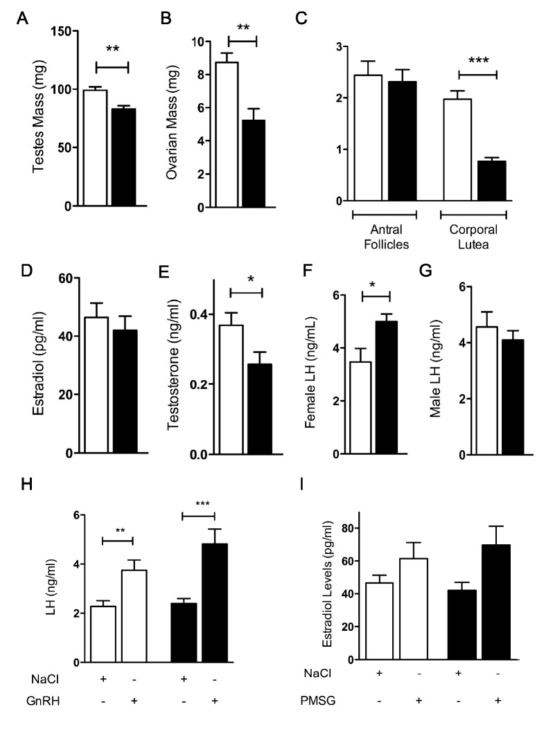
Figure 5. nes-Cre;Dmxl2 –/wt mice displayed a partial gonadotropin deficiency.
(A and B) Weights of testes and ovaries were low in nes-Cre;Dmxl2 –/wt mice. (C) Histological analysis of ovaries showed a normal number of antral follicles but very few corpora lutea in female nes-Cre;Dmxl2 –/wt mice. (D) Estradiol concentrations were normal in female nes-Cre;Dmxl2 –/wt mice. (E) Plasma testosterone concentration was low in male nes-Cre;Dmxl2 –/wt mice. (F) Plasma LH concentrations were moderately high in female nes-Cre;Dmxl2 –/wt mice. (G) Despite their lower testosterone concentrations, male nes-Cre;Dmxl2 –/wt mice had normal plasma LH concentrations. (H) The GnRH-induced increase in LH concentration was normal in nes-Cre;Dmxl2 –/wt mice. (I) The administration of PMSG to young mice induced a normal increase in estradiol concentration in nes-Cre;Dmxl2 –/wt mice, similar to that observed in their WT littermates (asterisks indicate significant differences: * p<0.05, ** p<0.001; ***p<0.0001). Error bars: SEM. P, postnatal day. White bars, Dmxl2lox/wt; black bars, nes-Cre;Dmxl2 –/wt. Numerical data used to generate these graphs may be found in Table S5.

