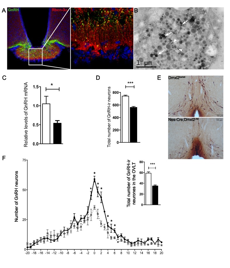
Figure 6. Hypothalamic GnRH mRNA and GnRH-IR neuron levels are lower in the hypothalamus of nes-Cre;Dmxl2 –/wt mice.
(A) Rbcn-3α is expressed in GnRH neurons in the median eminence. (B) Rbcn-3α is located in small clear vesicles and in LDCVs in GnRH neurons. White arrows indicate Rbcn-3α DAB staining; white arrow heads indicate GnRH nanogold staining. (C) GnRH1 mRNA levels relative to RNA18S were lower in the hypothalamus of nes-Cre;Dmxl2 –/wt male mice than in WT mice. (D) The total number of GnRH-ir neurons in the brain was lower in nes-Cre;Dmxl2 –/wt male mice than in WT mice. (E) GnRH immunostaining in the OVLTs in Dmxl2 lox/wt and nes-Cre;Dmxl2 –/wt male mice. (F) An analysis of the rostral–caudal distribution of GnRH-ir neurons in the hypothalamus revealed that nes-Cre;Dmxl2 –/wt male mice had fewer GnRH-ir cell bodies in the OVLT (see inset) than their WT littermates. * p<0.05, *** p<0.0001. White bars, Dmxl2lox/wt; black bars, nes-Cre;Dmxl2 –/wt. Numerical data used to generate graphs 6C, 6D, 6F, and 6F inset may be found in Table S5.

