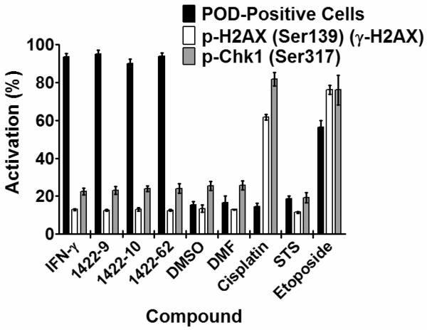
Figure 4. POD-inducing N-methyl triamines do not induce DNA damage.
HeLa cells were treated for 12 h with IFN-γ (4 U/μL), DMSO (0.1%), DMF (0.1%), cisplatin (25 μM), staurosporine (25 nM), etoposide (25 μM), or 1422 compounds (10 μM). Plates were then immunostained for PODs (mouse monoclonal anti-human PML, Alexa Fluor 488 chicken anti-mouse antibodies), phospho-H2AX (rabbit polyclonal anti-human phospho-H2AX-Ser139, Alexa Fluor 568 goat anti-rabbit antibodies), or phospho-Chk1 (rabbit polyclonal anti-human phospho-Chk1-Ser317, Alexa Fluor 568 goat anti-rabbit antibodies). POD positive nuclei (%) were quantified as previously described. p-H2AX and p-Chk1 nuclear staining intensity was quantified using CytoShop. Mean and standard deviation are shown (n=4 wells/condition, >200 cells imaged and quantified per well).

