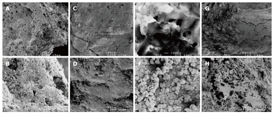
Figure 2.

Scanning electron microscopy images of some bone graft substitutes in the negative control group. A, B: Highly porous β-tricalcium phosphate (β-TCP) - (Negative control group); C, D: β-TCP (Negative control group); E, F: Hydroxyapatite (Negative control group); G, H: Calcium sulphate (Negative control group).
