
Figure 2.
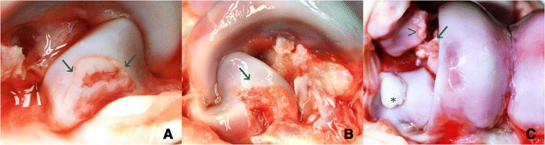
Osteochondrosis dissecans lesions in the coracoid process of calcaneus. A. Lateral close-up of the right coracoid process: multiple clefts in the articular cartilage expose underlying subchondral bone tissue and have caused synovitis with increased amounts of transparent/slightly haemorrhagic synovial fluid. B. Lateral view of the left coracoid process: a large fragment of articular cartilage has loosened from the subchondral bone tissue (arrow). The exposed bone and surrounding synovial and soft tissues are inflamed. C. Frontal view of the right lateral trochlea and coracoid process: osteochondral fragment (*) adherent on the surface of the coracoid process. Kissing lesions on the lateral malleolus of the fibula (arrowhead) and fascies articularis malleolaris of the talus (arrow) are also seen.