Figure 1.
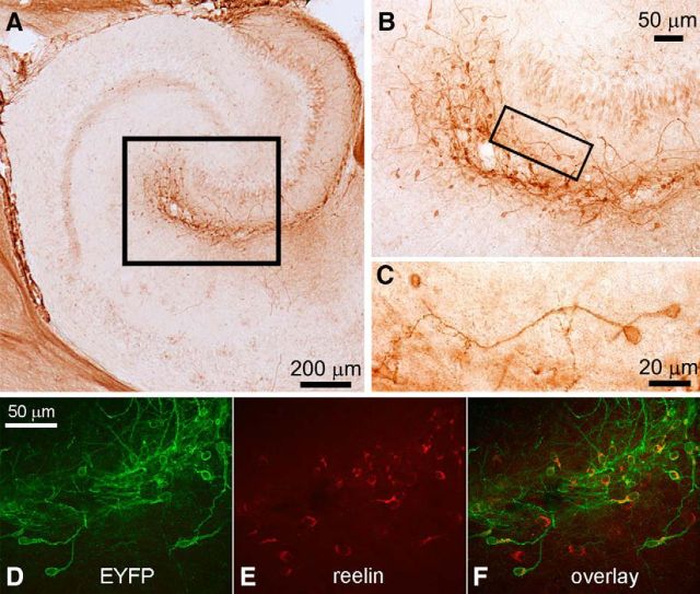
Bright-field and confocal microscopy demonstrate ChR expression in hippocampal Cajal-Retzius cells. Images from transverse hippocampal sections of P14 Wnt3a-IRES-Cre; ChR2(H134R)-EYFP animals. A, Immunocytochemistry shows EYFP-labeled cells along the hippocampal fissure in stratum lacunosum-moleculare and molecular layer of the dentate gyrus (black box). B, The area included in the box in A is shown at higher magnification. Notice the typical features of Cajal-Retzius cells. A black box selects an individual cell, which is shown at higher magnification in C. D–F, Confocal images showing the immunolocalization of EYFP, reelin, and their overlap.

