Figure 12.
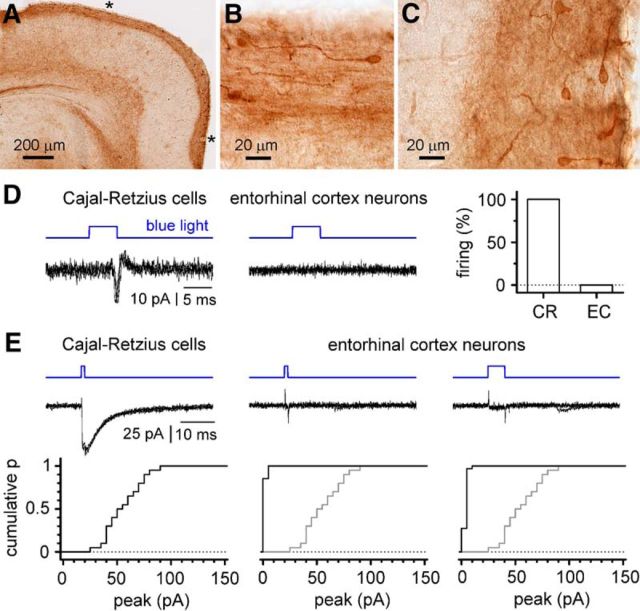
Lack of direct light-evoked firing and of ChR expression in entorhinal cortex neurons. A, Immunocytochemistry showing EYFP labeling of layer I in the entorhinal cortex, but no clearly labeled neurons in the deeper layers. B, C, Enlargements of the areas marked by an asterisk in A for the lateral and medial entorhinal cortices, respectively. Notice the typical morphology of Cajal-Retzius cells and the staining associated with layer I. D, Comparison of the response to light in hippocampal Cajal-Retzius neurons (left) versus layer II/III entorhinal cortex cells (middle) in cell-attached conditions. Notice the reliable firing reflected by action currents in Cajal-Retzius cells and the lack of light-induced effects in entorhinal cortex neurons. Four sweeps superimposed in both cases. Right, The percentage of cells responding to light with firing in the two different samples. CR, Cajal-Retzius cells; EC, entorhinal cortex neurons. Notice that entorhinal cortex neurons do not fire under these experimental conditions. E, ChR-mediated currents in hippocampal Cajal-Retzius cells (left), and layer II/III entorhinal cortex neurons (middle and right). A short flash of light (1 ms) produces significant currents in Cajal-Retzius cells, as shown by the recording (four traces superimposed, top) and by the cumulative distribution plot for several experiments (bottom). In contrast, light flashes either of the same (1 ms; middle) or of longer duration (5 ms; right) do not produce significant currents in entorhinal neurons. Top, Four traces superimposed showing the response of entorhinal cortex neurons to 1 and 5 ms flashes. Bottom, Summary plots for the two conditions (the distribution of the photocurrents in Cajal-Retzius cells is also shown in gray for comparison).

