Figure 2.

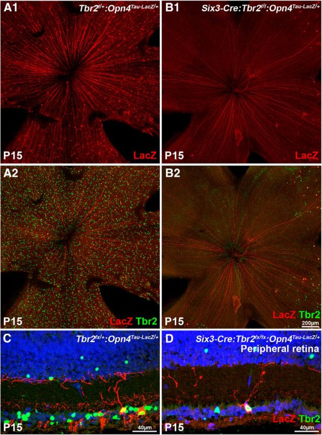
Tbr2 is essential for the formation of ipRGCs. Confocal images of P15 control and Tbr2-deficient retinal flat mounts (A, B) and cryosections at the peripheral region (C, D) that show Tbr2 and LacZ (reporting Opn4) labeling. A1, A2, C, The control Tbr2flox/+:Opn4Tau–LacZ/+ retinas show normal distribution of Tbr2+ and LacZ+ cells. A1 shows only the red channel from A2. B1, B2, D, The mutant Six3–Cre:Tbr2flox/flox:Opn4Tau–LacZ/+ retinas show the absence of Tbr2+ and LacZ+ cells in the central retina. Only a few LacZ+ cells can be found in the periphery. All remaining LacZ+ cells are also positive for Tbr2, indicating that Tbr2 is not removed by Cre in these cells. B1 shows only the red channel from B2.
