Figure 3.
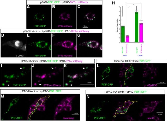
SYT-α puncta colocalize with neuropeptide-GFP puncta in cultured neurons. The cotransfection of dimm, PDF-GFP, and SYT-α-mCherry in the Drosophila neuronal BG3–c2 cell line. There is enhanced accumulation and colocalization of neuropeptide and SYT-α protein, dependent on DIMM expression. A–C, Representative levels of PDF-GFP (green) and SYT-α-mCherry (magenta) fusions without DIMM. D–G, The same but now with dimm cotransfection. H, Quantitative measurement of fluorescence intensity within the selected area indicates significant increases with DIMM cotransfection. **p < 0.01. I–K, Higher magnification of processes from a dimm-transfected cell showing correspondence of the two fluorescent signals indicating a subcellular association between puncta containing neuropeptide cargo and SYT-α (arrow). L–N, Representative labelings of PDF-GFP (green) and the intracellular markers (magenta), including two Golgi markers (dGM130 and lava lamp) (L, M) and the ER exit site marker (Sec16, N).

