Figure 4.

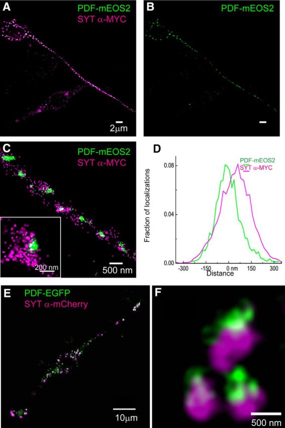
Coassociation of SYT-α and neuropeptide cargo with LDCV-like vesicles in cultured Drosophila neurons. STORM/PALM image of a Drosophila neuronal BG3–c2 cell transiently transfected with plasmids encoding DIMM (data not shown), PDF-mEOS2, and SYT-α-MYC (A–C). Punctate clusters of PDF-mEOS2 are distributed throughout the neuronal process (B), where they appear highly coassociated with SYT-a-MYC localizations. Zoomed-in images indicate a structure with LDCV-like dimensions, wherein SYT-α-MYC localizations decorate PDF-mEOS2 clusters (C). Frequently, SYT-α-MYC localizations appear polarized adjacent to those of PDF-mEOS2 (C, inset). A projection histogram of these localizations obtained from ∼50 aligned clusters. D, Quantitative analysis represents this polarized localization pattern. SIM images of a BG3–c2 cell coexpressing DIMM, PDF-eGFP, and SYT-α-mCherry indicate a similar polarized association of SYT-α-MYC adjacent to cargo (E, F).
